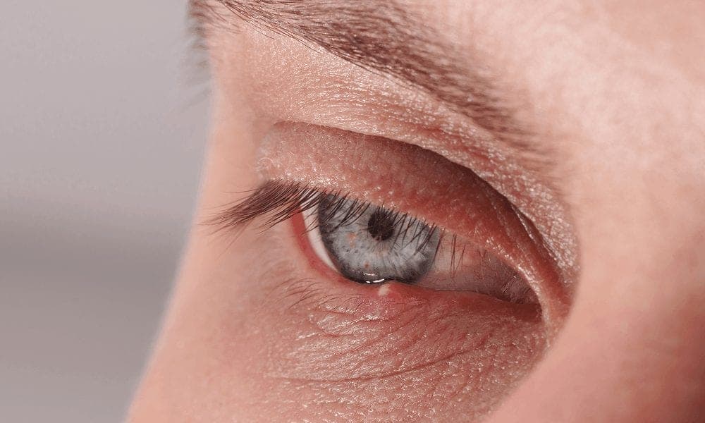
Our eyelids are more than just delicate folds of skin; they act as protective shields, natural moisturisers, and even play a role in the expressiveness of our eyes. From protecting the eyeball against dust and light to spreading tears evenly across the eye’s surface, eyelid anatomy is fascinating and essential for healthy vision.
But what exactly makes up the eyelid? In this blog, we’ll explore the anatomy of the upper eyelid, the lower eyelid, and parts of the outer eyelid in detail. By the end, you’ll understand how these small yet complex structures work together to keep your eyes safe and healthy.
Movement -
Eyelid Anatomy Overview
Functions of the Eyelid
The main functions of the eyelid anatomy include:- Protection - shielding the eyes from injury, bright light, and dust.
- Moisturising - spreading tears across the cornea to prevent dryness.
- Barrier - reducing infection risk by blocking foreign particles.
Parts of the Eyelids
The upper lid anatomy and lower lid anatomy are made up of several essential components that work together to protect the eye and maintain its function. These include:- Skin - The outermost layer, providing protection.
- Eyelashes - Act as a natural barrier against dust and debris.
- Conjunctiva - A transparent layer that keeps the eye moist.
- Orbicularis Oculi Muscle - Controls eyelid movement and blinking.
- Tarsal Plates - Provide structural support to the eyelids.
- Meibomian Glands - Produce an oily substance to prevent tear evaporation.
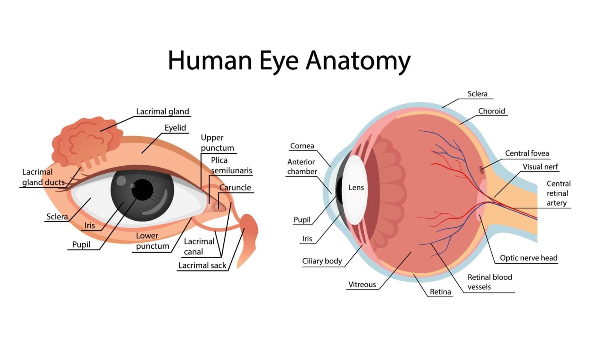
- Lacrimal Glands - Responsible for tear production.
- Nerves - Control movement and sensation in the eyelids.
- Extraocular Muscles - Help in coordinated eye movement.
- Fat Pads - Provide cushioning and support to the eyelids.
Key Parts of the Eyelid
1. Skin and Eyelashes
The eyelid skin is the thinnest in the human body, which is why swelling from fluid build-up or allergies appears so quickly here. Eyelashes act as tiny protective sensors. They block dust and irritants and instantly trigger a reflex blink when something comes too close. Alongside them, the glands of Zeis (oil) and Moll (sweat) help keep the lash line healthy. Fun fact: Eyelashes fall out naturally every few weeks and regrow within 6-8 weeks.2. Conjunctiva: The Invisible Lubrication Layer
The conjunctiva is a clear membrane that lines the inner eyelid and covers the white of the eye. It ensures constant lubrication, comfort, and infection prevention.- Palpebral conjunctiva - lines the inner eyelid.
- Tarsal conjunctiva - directly attached to the tarsal plate.
- Bulbar conjunctiva - covers the white part of the eye.
3. Orbicularis Oculi Muscle
This ring-shaped muscle controls eyelid closure. It has two main parts:- Palpebral portion - responsible for gentle blinking.
- Orbital portion - allows tight eyelid closure, such as when squinting.
4. Glands
The eyelid structure includes several glands that ensure proper lubrication and tear production. Glands in our body release substances that help it work well. In our eyes, there are special glands that release liquids to keep our eyes moist and comfortable. Meibomian glands make oil to keep our eyes lubricated, while others release sweat to cool them down. The glands in our eyelids also produce oil for our eyelashes. When we cry, most of our tears come from the lacrimal glands above our eyes and then spread across the front of our eyes. The extra tears are drained into our noses, which is why our noses run when we cry.5. Nerves - Connectors for Movement and Sensation
It really requires a lot of control to make your eyes blink correctly. These control our eye movement and feelings:Movement -
- Three types of nerves send signals from the centre of the body that help the eyelids move.
- The seventh cranial nerve, also called the facial nerve.
- The third cranial nerve, also called the oculomotor nerve.
- Sympathetic nerves, which make the eyelids move without us thinking about it.
6. Extraocular Muscles
The anatomy of the eyelid is incomplete without mentioning the extraocular muscles that control eye movement. Six muscles on the outside of the eye help it move around. These muscles can move the eye up, down, and to the sides. Together, they can shift the eye in any direction.- Medial Rectus (MR) - moves the eye towards the nose.
- Lateral Rectus (LR) - displaces the eye from the nose.
- Superior Rectus (SR) - moves the eye upward and inward.
- Inferior Rectus (IR) - moves the eye inward and downward.
- Superior Oblique (SO) - moves the eye outward and downward.
- Inferior Oblique (IR) - moves the eye upward and outward.
7. Fat Pads
In and around the eyelids, there are different layers of fat. One layer of fat is found behind the orbital septum and in front of the levator aponeurosis. In the upper eyelid, there are two more fat pads located in the middle and towards the nose. The lower eyelid fat pads have a slightly different structure. The central fat pad is separated from the medial fat pad by the inferior oblique muscle. There is also a small amount of fat in front of the inferior oblique muscle. The inferior oblique muscle starts from the lower border of the orbital floor and moves behind the orbital margin and towards the upper aspect of the nasal lacrimal canal. It then goes underneath the inferior rectus muscle and through the Tenon capsule, ultimately getting close to the macula of the eye. Fat repositioning or removal is commonly performed during blepharoplasty to achieve a more youthful appearance.
Trust your eyes to the experts
- Consult with our doctor-led team for expert, personalised care
- A Care Quality Commission (CQC) rated clinic for your peace of mind
- Achieve a naturally refreshed look with over 25 years of expertise
Upper Eyelid Anatomy vs. Lower Eyelid Anatomy
- Upper Eyelid Anatomy: Contains the levator palpebrae superioris muscle, which helps lift the eyelid, and a larger tarsal plate for structure. It plays a critical role in vision, as drooping can obstruct sight.
- Lower Eyelid Anatomy: Has a smaller tarsal plate and is more prone to puffiness due to fat pads and fluid retention. It doesn’t move as much as the upper lid, but it helps with tear drainage and protection.
- Outer Eyelid Parts: The corners of the eyelids (medial and lateral canthus) are essential in maintaining eyelid alignment and tear flow.
Common Eyelid Conditions Related to Anatomy
Understanding eyelid anatomy also helps in recognising common issues such as:- Ptosis - drooping upper eyelid due to muscle or nerve weakness.
- Blepharitis - inflammation of the eyelid margin.
- Chalazion or stye - blockage of Meibomian or oil glands.
- Eyelid malpositions (entropion, ectropion) - where the eyelid turns inward or outward.
Conclusion
The eyelid anatomy may look simple, but it is made up of complex structures. From the parts of the outer eyelid that align the lids to the fat pads, glands, and muscles within the upper lid anatomy and lower lid anatomy, each part has a vital role in vision and protection. By understanding the anatomy of the upper eyelid and the anatomy of the lower eyelid, we can better appreciate how these structures maintain healthy eyes.Good to know
Frequently asked questions
Still unsure? Our team is glad to talk you through anything before you book.
- Your eyelids have tiny oil glands called Meibomian glands, which sit along the edge of your eyelids near your eyelashes. These glands produce an oily substance that helps keep your tears from drying out too quickly. If these glands get blocked or inflamed, it can lead to eye problems like dryness or irritation.
- Your eyelids act like protective covers for your eyes, shielding them from dust, bright light, and other outside elements. They also help keep your eyes moist by spreading tears evenly across the surface. Unlike other parts of your skin, eyelids don’t have fat underneath, making them thinner and more delicate.
- Eyelids come in different shapes, giving everyone a unique look. The most common types include: Double eyelids - Have a natural crease that makes the upper eyelid fold.; Monolids - Lack a visible crease, giving the eyelid a smooth appearance.; Hooded eyes - Have extra skin that folds over the crease, sometimes making the eyes look smaller.
- A lazy eyelid is medically known as ptosis or blepharoptosis. This condition causes one or both upper eyelids to droop. In some cases, the drooping is mild, but in severe cases, it can cover part of the pupil and block vision.
- If you have a swollen or blocked oil gland (like a chalazion), here are some simple home treatments: Warm compress: Soak a clean cloth in warm water and hold it over the swollen area for about 15 minutes, at least three times a day. This helps open the gland and drain any blockage.; Good hygiene: Avoid wearing eye makeup until the swelling goes away, and keep the area clean even after it drains.